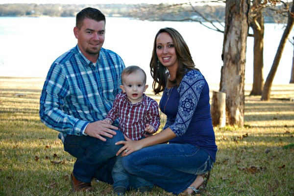
Watching Jaxon play with monster trucks is truly a precious miracle!

A week overdue for labor and delivery of their first child, Alvaradians Cody and Carly Bearden were taken by surprise when they learned through the sonogram, given as a precaution prior to inducing labor at Cooks Children Hospital Fort Worth, that their baby boy, Jaxon Bearden, had something wrong with his heart. This was not an immediate finding. In fact, the first review of the sonogram invoked reactions that were of low concern.
“The medical staff told us that they noticed he had a little problem, but after he was born, they performed an echocardiogram (EKG) and found that he had an aortic coarctation, a critical congenital heart defect” said Carly. “We were hoping for the best, and we were not prepared for that news – it was a complete shock.” Attending cardiothoracic surgeon, Vincent K.H. Tam, MD, and pediatric cardiologist, Matthew Dzurik, MD, suggested that the open heart surgery be performed when Jaxon was five days old because all of his vital signs were very good.
“The days leading up to Jaxon’s surgery were both calming and stressful, because the doctors and staff made us feel so assured with examples of the success rates with this type of surgery,” explained Carly. “A chaplain came to pray with us and a hospital counselor came by to add to our comfort level.”
When Jaxon was still in the neonatal intensive care unit (NICU), a special care package was given to the Beardens by a family whose son did not survive his heart condition, The giving is a form of legacy that the family of the deceased provides as a gift to all little patients undergoing cardiac procedures within this NICU at Cooks Children Hospital Fort Worth. Mrs. Bearden discussed that Jaxon was only in the hospital for a total of 11 days, six days after his surgery, because the doctors were so impressed with his recovery status. The family was (and is) allowed to carry out their lives as normally as any family of a newborn would without such a complication. There were no concerns and only minimal warnings such as: becoming short of breath, irregular breathing, abnormal eating habits, and disruptions in sleep behaviors because the doctors really did not expect these to occur. No monitors or special equipment were needed.
Jaxon went for a checkup two weeks after his release and was seen quarterly for a year. Although his checkups are now yearly, when he reaches age 13-18, the cardiologist will revisit the monitoring as he begins to play sports. To date, no other occurrences have taken place. To see him play as shown in the photos below, getting excited about watching and playing with monster trucks … Gravedigger, specifically, and high-fiving his mother as he shows off his truck is truly a precious miracle!
“Sometimes we forget that Jaxon had heart complications. He seems like an average healthy baby,” Carly boastfully exclaimed. “Nothing would have raised a flag without the sonogram that was taken when Jaxon was late – the anatomy scan showed no flags during my pregnancy.” However, she quickly pointed out that this sonogram was part of the wonderful standard of care provided by the physicians. She mentioned learning that every baby is born with a patent ductus arteriosus (ductus arterious [PDA]), a small blood vessel that connects the aorta and the main pulmonary artery. The ductus arterious allows blood to bypass the lungs during pregnancy because the baby gets air from the mother.
According to the Centers of Disease Control and Prevention (CDC), the defect occurs when a baby’s aorta does not form correctly as the baby grows and develops during pregnancy. The narrowing of the aorta usually happens in the part of the blood vessel just after the arteries branch off to take blood to the head and arms, near the PDA, although sometimes the narrowing occurs before or after the ductus arteriosus. In some babies with coarctation, it is thought that some tissue from the wall of ductus arteriosus blends into the tissue of the aorta. When the tissue tightens and allows the ductus arteriosus to close normally after birth, this extra tissue may also tighten and narrow the aorta. The narrowing, or coarctation, blocks normal blood flow to the body. This can back up flow into the left ventricle of the heart, making the muscles in this ventricle work harder to get blood out of the heart. Since the narrowing of the aorta is usually located after arteries branch to the upper body, coarctation in this region can lead to normal or high blood pressure and pulsing of blood in the head and arms and low blood pressure and weak pulses in the legs and lower body. If the condition is very severe, enough blood may not be able to get through to the lower body. The extra work on the heart can cause the walls of the heart to become thicker in order to pump harder. This eventually weakens the heart muscle. If the aorta is not widened, the heart may weaken enough that it leads to heart failure. The Centers for Disease Control and Prevention (CDC) estimates that about 4 out of every 10,000 babies are born each year in the United States with coarctation of the aorta.1
“I wish I could educate more new mothers of the risks of not going for all of their prenatal visits during their pregnancy,” proclaimed Carly. “Prenatal visits and sonograms are so important, and again, had they not took the sonogram when Jaxon was a week late on his predicted birth date, we would not have been so lucky.”
Carly hopes that by reading this story someone else is helped and another life is saved.
– Carly Bearden, Alvarado, Texas
